Introduction
Understanding the electrical axis of the heart through an electrocardiogram (ECG) is crucial for diagnosing various cardiac conditions. Calculating the heart axis from an ECG involves analyzing the direction and magnitude of the electrical activity perceived during the cardiac cycle. This webpage provides an in-depth guide on how to calculate axis in an ECG, which is often a pivotal part of cardiac health assessments.
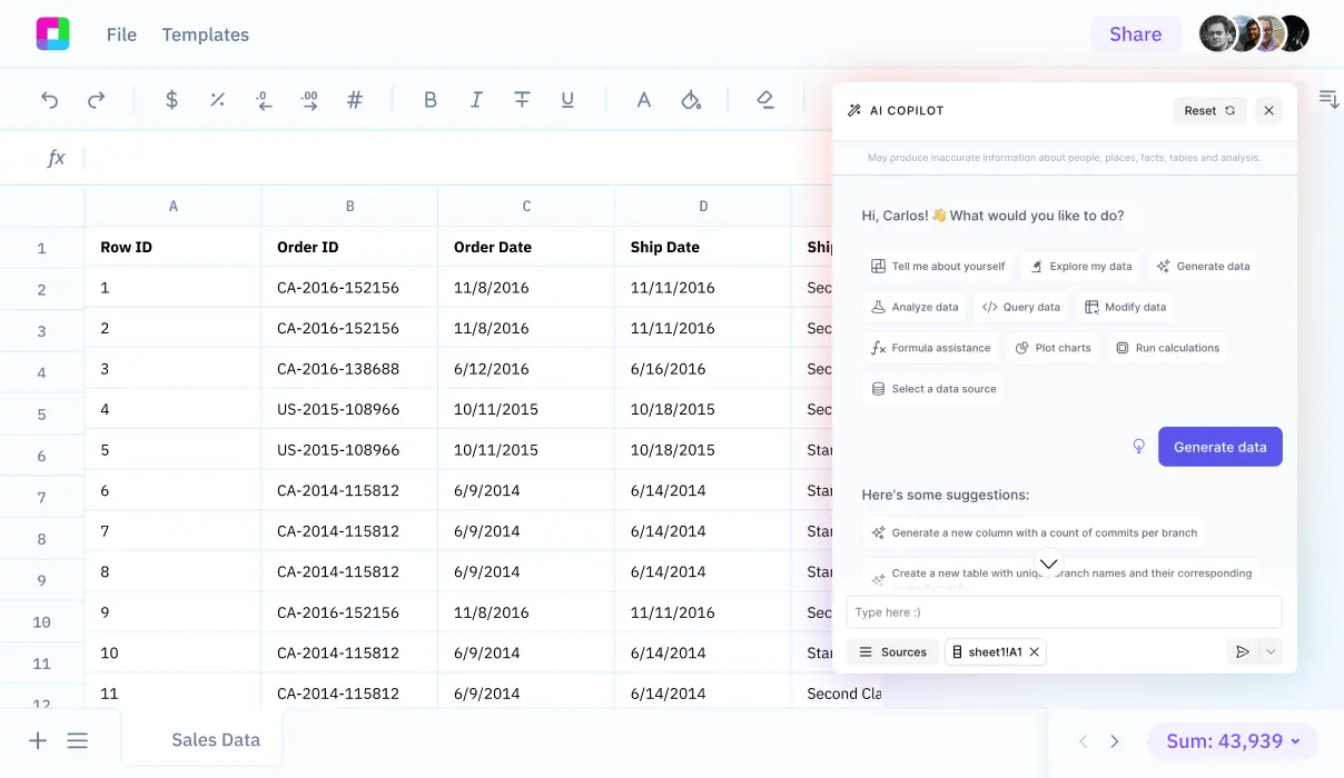
We will additionally delve into how Sourcetable simplifies this calculation with its innovative AI-powered spreadsheet assistant. This tool helps automate intricate calculations and enhance the accuracy of your analyses. Experience the convenience and precision of Sourcetable by signing up at app.sourcetable.cloud/signup.
How to Calculate Axis in ECG
Determining the heart axis in an electrocardiogram (ECG) involves analyzing the direction of the ventricular depolarization vector. This is critical for diagnosing conditions related to the cardiac axis deviation.
Tools Needed for ECG Axis Calculation
To calculate the ECG axis, specific leads are necessary depending on the method used. The Quadrant Method requires leads I and aVF. The Three Lead analysis, a more detailed approach, uses leads I, II, and aVF. For those employing the Isoelectric Lead analysis, any ECG lead that shows an isoelectric QRS can be utilized. A versatile method, Super SAM the Axis Man, uses multiple leads to determine the axis accurately.
Steps to Calculate Axis in ECG
The process begins with the selection of the appropriate method and corresponding leads. Simple methods primarily use leads I and aVF to ascertain if the heart axis is normal or deviated. The more accurate method involves identifying a biphasic lead and using it along with leads I and III. Measurements from these leads are then translated into the hexaxial reference system for precise axis determination.
Understanding Axis Deviations
The normal cardiac axis ranges between -30° and +90°. An axis less than -30° indicates a left axis deviation, while an axis greater than +90° points to a right axis deviation. Extremes, where the axis ranges between -90° and +180°, are categorized as indeterminate or extreme deviations. These deviations suggest various underlying cardiac conditions, emphasizing the importance of precise axis calculation.
Special Considerations in Pediatric ECG
In pediatric ECG interpretation, the normal axis ranges from +30° to +190° at birth, gradually moving leftward as the child ages. This shift underlines the importance of age-specific axis consideration in pediatric cardiology.
Understanding and applying the appropriate method using the correct tools are essential in accurately calculating the ECG axis, which is pivotal in diagnosing and managing various cardiac anomalies effectively.
How to Calculate Axis in ECG
Understanding ECG Axis
The ECG axis represents the predominant direction of heart's electrical activity. Accurate determination of the QRS axis, the most critical, influences diagnosis and treatment decisions. Normal, left, and right axis deviations offer insights into cardiac condition.
ECG Axis Calculation Methods
Three straightforward methods facilitate the estimation of the electrical heart axis: the Quadrant Method, the Three Lead Method, and the Isoelectric Lead Method.
The Quadrant Method
Use Lead I and aVF. If the QRS complex is positive in both, it indicates a normal axis (0° to +90°). A positive QRS in Lead I and negative in aVF suggests left axis deviation (LAD) while the opposite indicates right axis deviation (RAD).
The Three Lead Method
Extends the Quadrant approach by incorporating Lead II. This method refines the axis estimation, especially useful to identify an indeterminate or extreme axis, positioned between +/-180° and -90°.
The Isoelectric Lead Method
Identifies the isoelectric lead, where the QRS complex shows no net amplitude. Then, analyze adjacent leads to determine the positive quadrant, aiding precise axis calculation. This method is valuable when the QRS axis is difficult to interpret through usual methods.
Step-by-Step Calculation
To determine the QRS axis, analyze the limb leads on the ECG. A normal axis falls between -30° and +90°. Deviations are classified based on the direction and amplitude of the QRS in specific leads, impacting the diagnostic interpretation significantly.
Examples and Importance
For example, a QRS that is positive in Lead I, II, and aVF typically indicates a normal axis. However, variations such as a negative QRS in Lead I and positive in aVF suggest RAD. Recognizing these patterns is crucial for accurate cardiac assessment.
Understanding how to calculate the ECG axis empowers healthcare professionals to detect potential heart issues effectively, improving patient outcomes through more tailored and timely medical interventions.
How to Calculate ECG Axis - Practical Examples
Example 1: Normal Axis Determination
To identify a normal heart axis in an ECG, check the QRS complex in leads I and II. A normal axis suggests the QRS complex is positive in both leads. Calculate the net direction of electrical heart activity; if it lies between -30° and +90°, the axis is normal.
Example 2: Left Axis Deviation
For left axis deviation, examine the QRS complex in lead I to be positive and in lead II to be negative. This deviation usually indicates an electrical axis between -30° and -90°. Left axis deviation often necessitates further investigation for underlying conditions like left ventricular hypertrophy.
Example 3: Right Axis Deviation
Right axis deviation can be identified when the QRS complex is negative in lead I and positive in lead II. This indicates an electrical heart axis beyond +90°. Conditions such as pulmonary disease or right ventricular hypertrophy might lead to right axis deviation.
Example 4: Extreme Axis Deviation
Known also as "Northwest Axis", extreme axis deviation occurs when the QRS axis is between -90° and +180°. This rare occurrence often requires prompt medical assessment for potential severe cardiopulmonary conditions.
Master Complex Calculations with Sourcetable
Sourcetable, the AI-powered spreadsheet, revolutionizes the way you handle calculations, especially in complex domains such as ECG axis determination. Whether for academic, professional, or personal growth, Sourcetable ensures precision and ease.
Calculate ECG Axis Flawlessly
Determining the ECG axis is critical for diagnosing heart conditions correctly. Sourcetable simplifies this with its AI assistant, which not only performs calculations but also explains them. Just query, "how to calculate axis in ECG," and watch the AI work through the steps in both spreadsheet and chat formats.
The AI assistant uses Leads I and Leads II readings to compute the axis, employing standard cardiac axis formulas. Visualization and interpretation are straightforward, accompanying each step with clear explanations.
Embrace Sourcetable for its ability to demystify complex calculations and elevate your understanding, making it invaluable for studies or professional healthcare settings.
Use Cases for Calculating ECG Axis
1. Diagnosis of Cardiac Conditions |
Identifying the electrical axis through the ECG allows for the diagnosis of various cardiac conditions such as right or left ventricular hypertrophy, bundle branch blocks, and congenital heart defects. |
2. Monitoring Treatment Effects |
ECG axis calculation plays a critical role in monitoring the effectiveness of treatments for conditions that alter cardiac axis, such as hypertension and myocardial infarction. |
3. Enhancing Risk Stratification |
Accurate ECG axis determination helps in risk stratification in patients with heart disease, guiding further diagnostic testing and treatment options. |
4. Supporting Clinical Decisions |
A well-calculated ECG axis aids clinicians in making informed decisions regarding patient management in emergency and non-emergency situations. |
5. Educational Tool |
Teaching healthcare professionals how to calculate ECG axis equips them with essential skills for interpreting ECGs accurately, enhancing overall patient care. |
Frequently Asked Questions
What are the common methods used to calculate the QRS axis in an ECG?
The common methods include the Quadrant Method using Lead I and aVF, the Three Lead Analysis using Lead I, Lead II, and aVF, and the Isoelectric Lead Analysis which involves determining the isoelectric lead.
How does the Quadrant Method work for calculating ECG axis?
The Quadrant Method involves using Lead I and Lead aVF to determine the axis. Depending on the positivity or negativity of these leads, the axis can be identified within specific quadrants.
What is the Isoelectric Lead Analysis method for ECG axis determination?
The Isoelectric Lead Analysis identifies the isoelectric lead (which has a zero net amplitude, such as a biphasic or flat-line QRS) and calculates the QRS axis. The axis is estimated to be at 90 degrees to the isoelectric lead and points in the direction of the positive leads or opposite to the negative leads.
How can the Three Lead Analysis help in determining the ECG axis?
This method uses leads I, II, and aVF to analyze the ECG. The axis is estimated by either observing the area of overlap between the colored areas from leads I and II, or by using the leads that show the tallest R waves to determine the direction of the heart's electrical axis.
Conclusion
Understanding how to calculate the axis on an ECG is crucial for accurate heart health assessment. This involves analyzing the electrical axis through Q, R, and S wave summations, which indicates the heart's electrical conduction path. Mastering this calculation is key for both healthcare professionals and students alike.
Try Sourcetable for Easier Calculations
For a streamlined calculation experience, Sourcetable offers a solution. As an AI-powered spreadsheet, Sourcetable simplifies the complexities involved in ECG axis calculations and more. This tool is particularly valuable for experimenting with AI-generated data, enhancing both learning and professional application.
Explore Sourcetable's capabilities today and discover a more efficient approach to medical calculations. Try it for free at app.sourcetable.cloud/signup.


